Pirop – Ophthalmic Scanner (A+B+CCT)
Echoson
Produkt
Pirop – Ophthalmic Scanner (A+B+CCT)
Pirop
PIROP – state-of-the-art ophthalmic ultrasonic device for Biometry ,

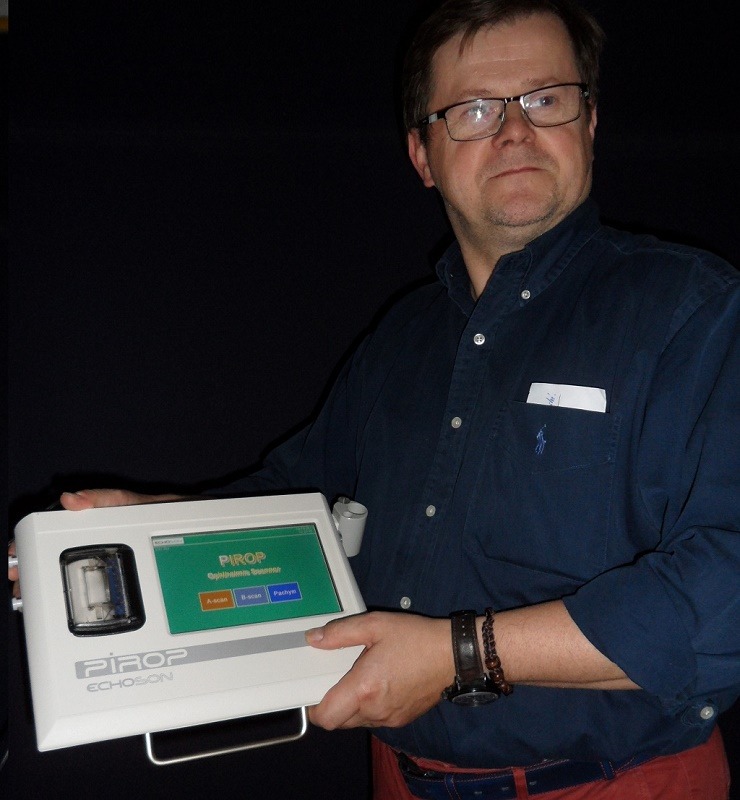
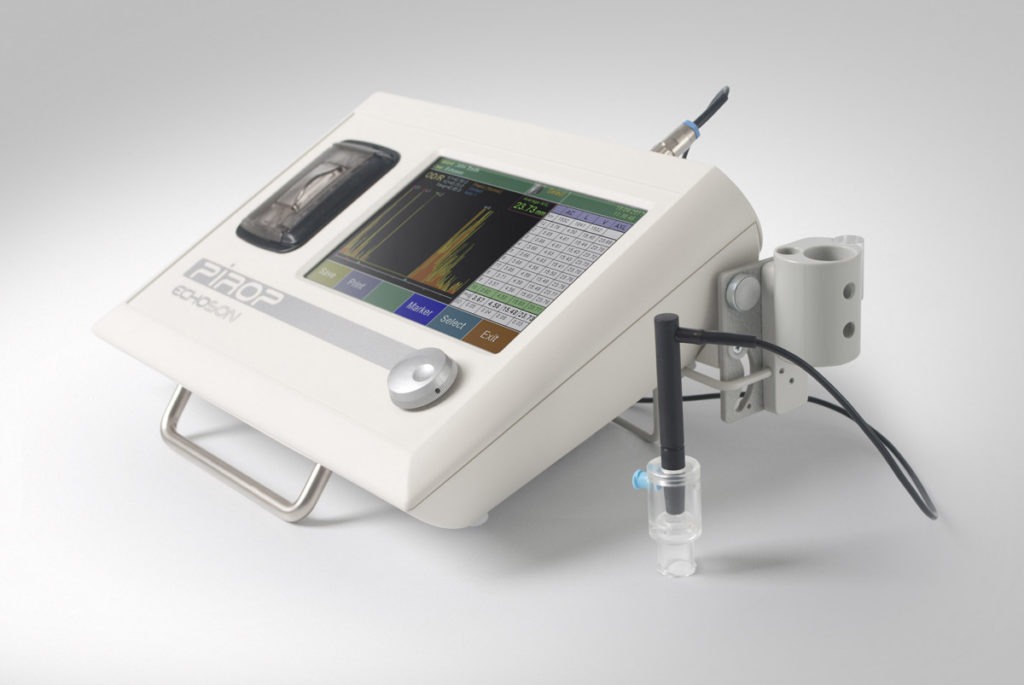
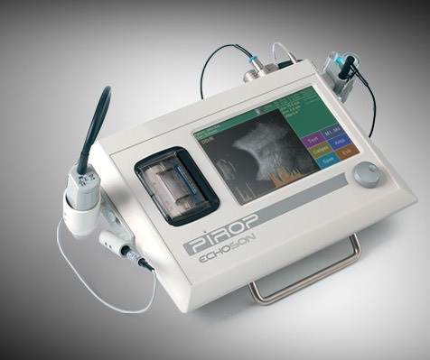
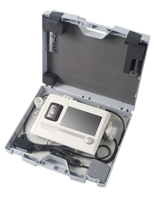
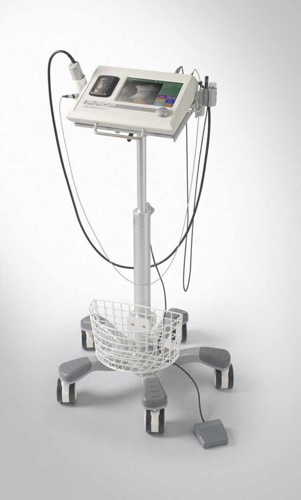
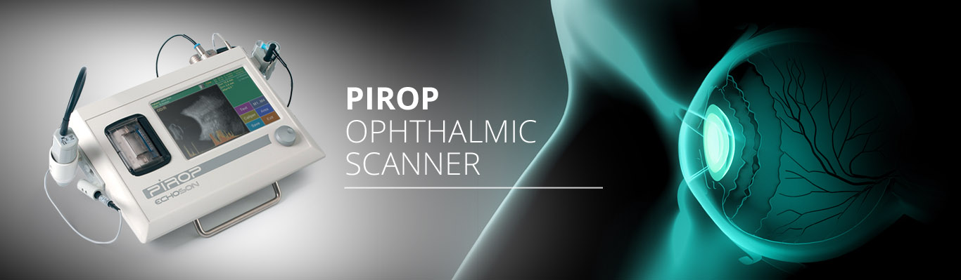
Visualization and Pachymetry of eyeballs   (A-Scan + B-Scan + CCT- Pachymeter) – modified version.

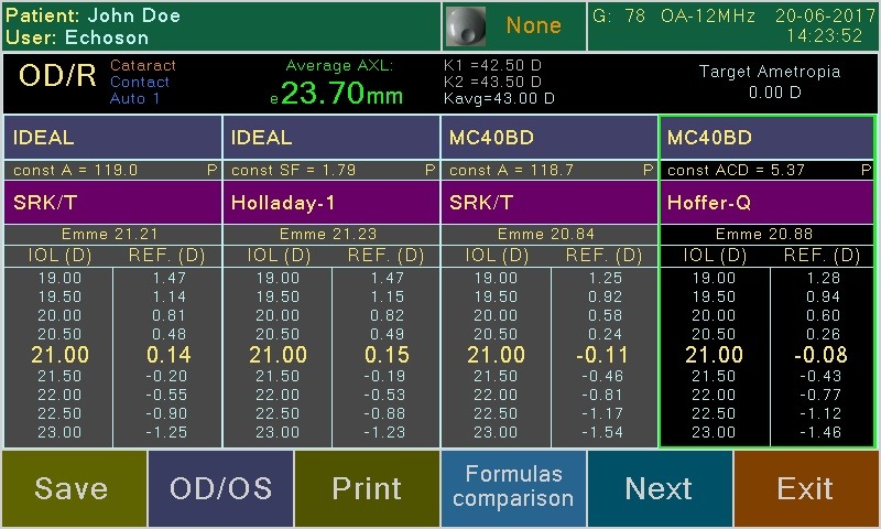
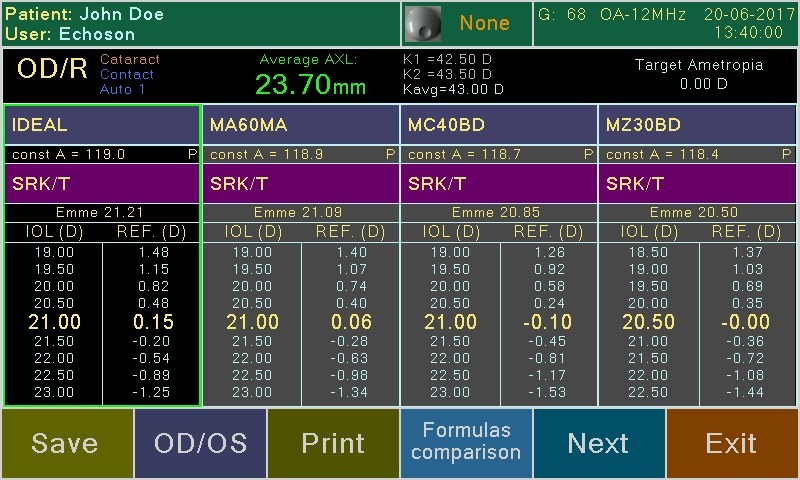
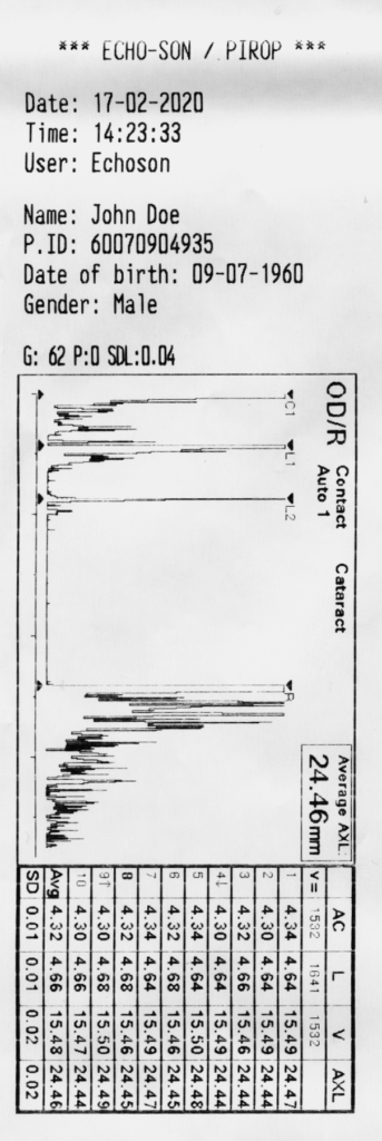
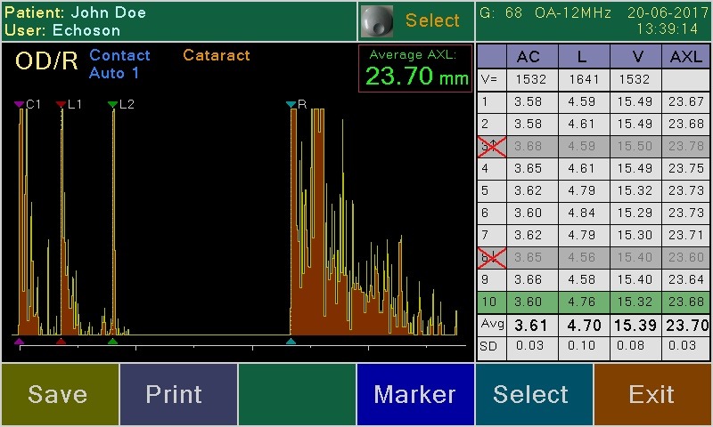
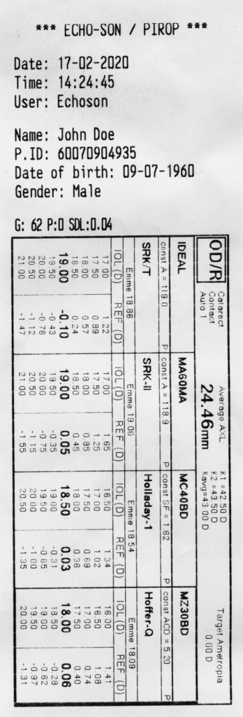
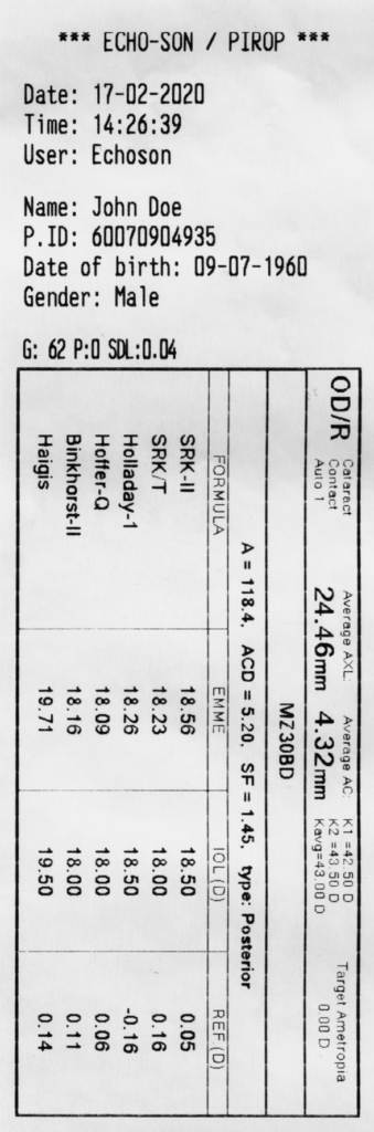
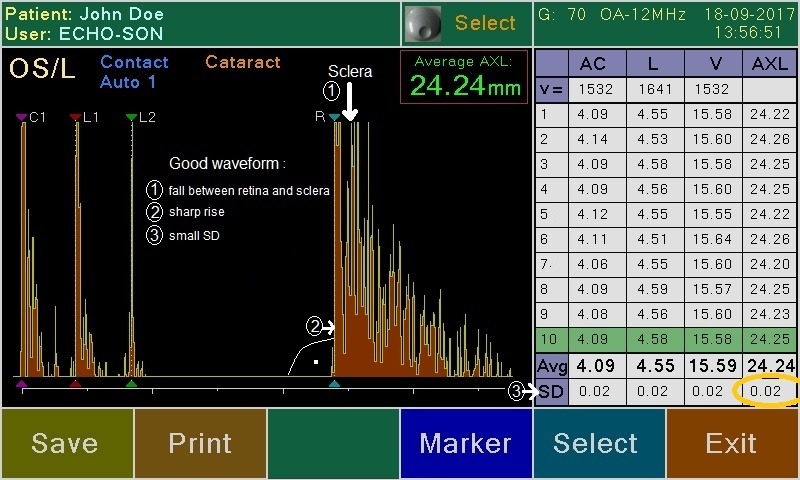
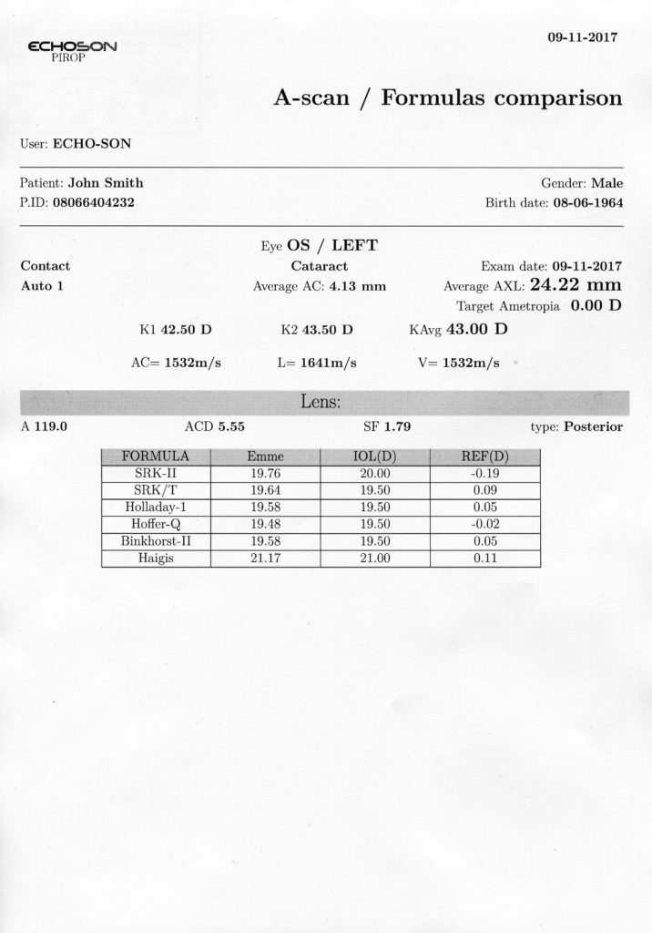
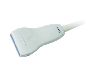
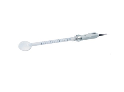
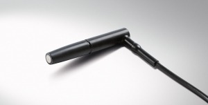
A-SCAN – Ocular Lens biometrics
Digital ‘A-scan’  tool for ophthalmology, biometry and lens power calculation of intra-ocular implants.

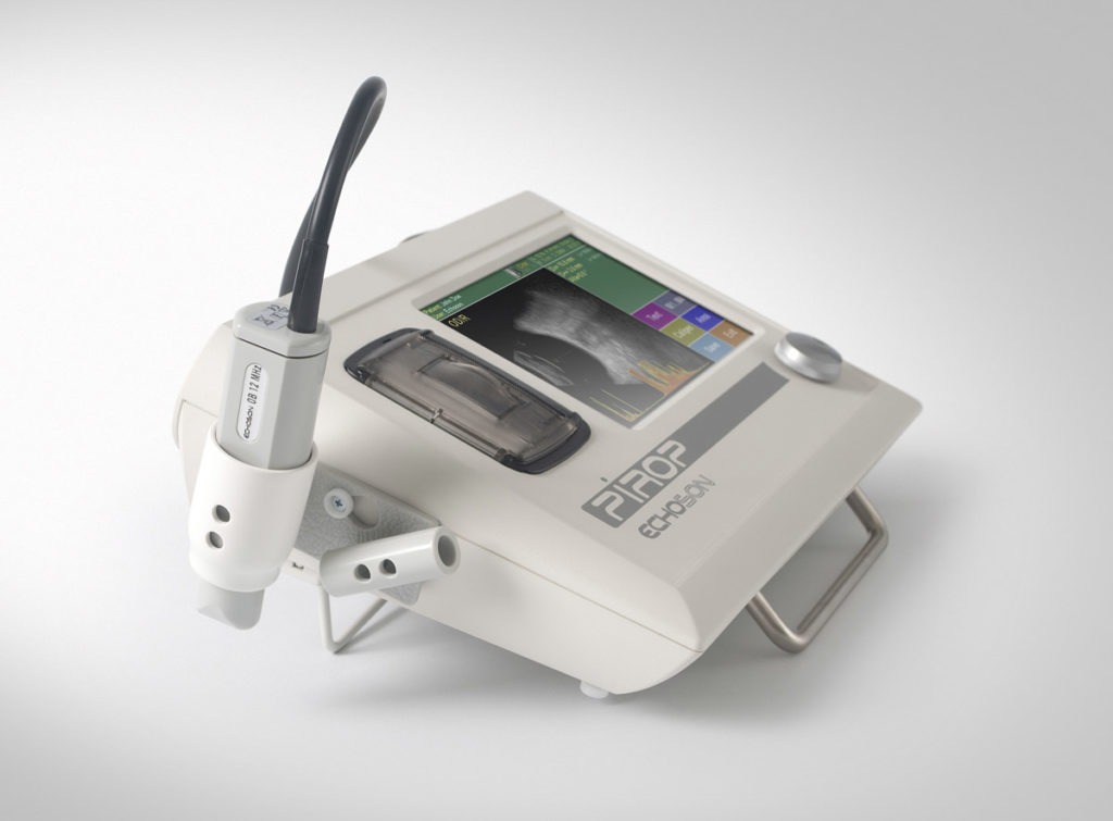
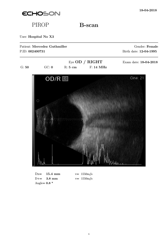
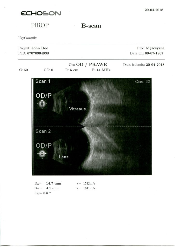
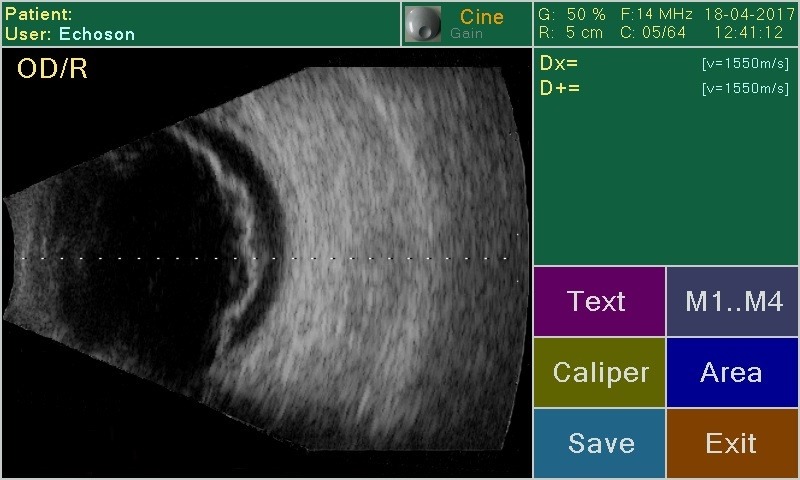
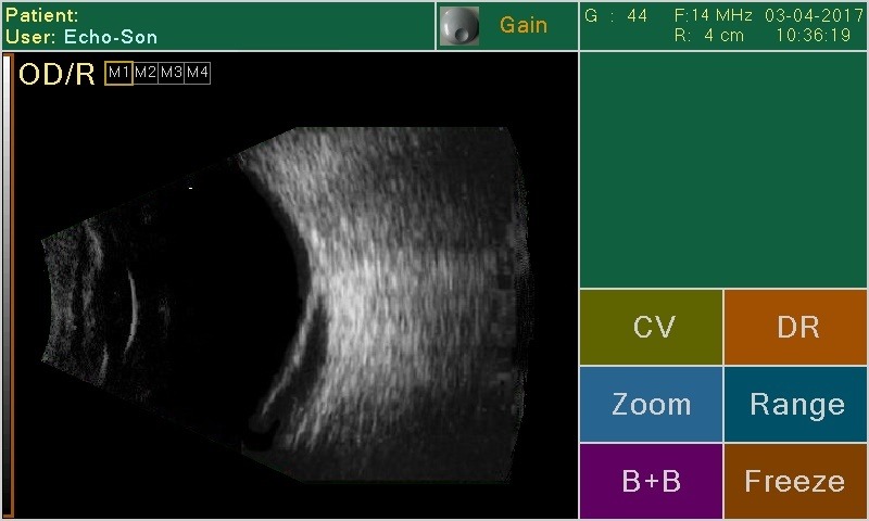
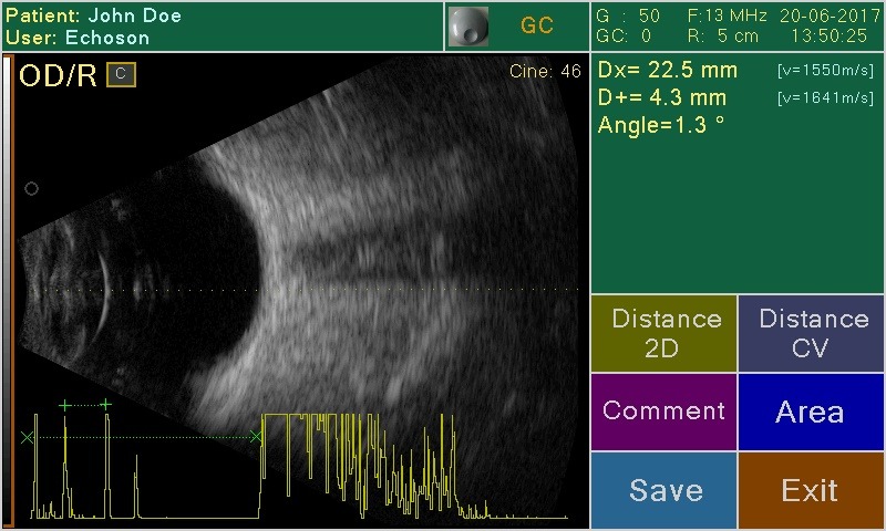
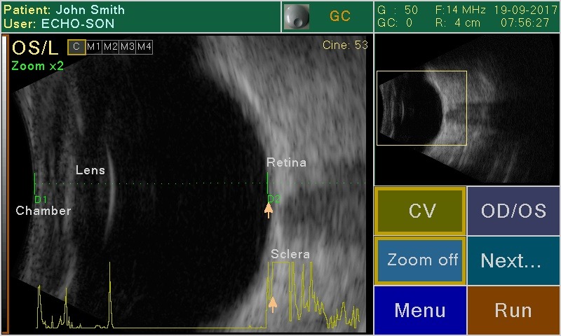
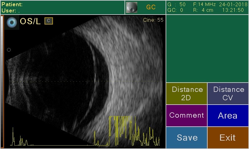
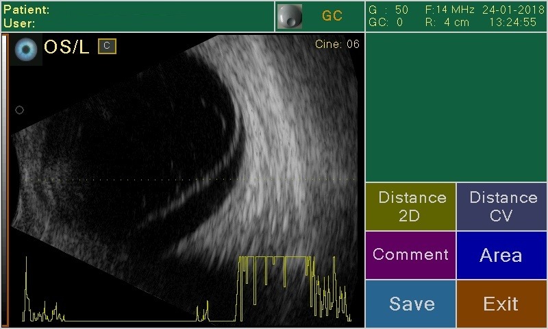
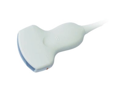
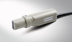
B-SCAN – Eye examination
Modern  ‘B-scan’  tool for ophthalmology,  interior eyeball imaging, retina, optical nerve …

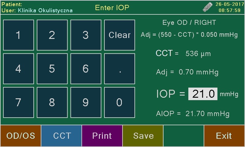
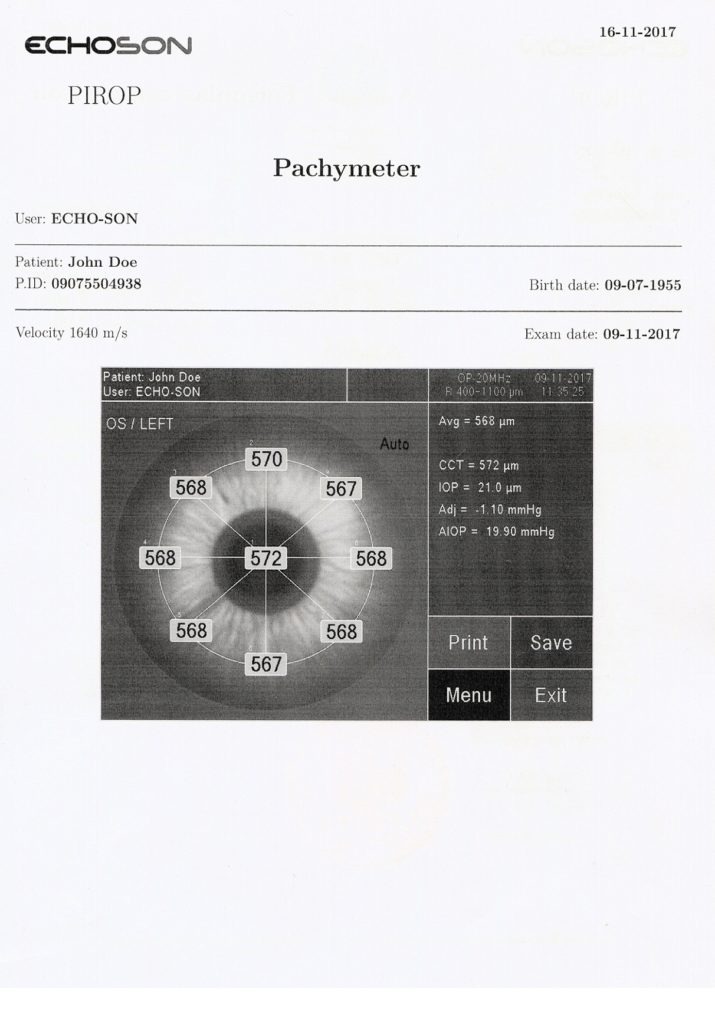
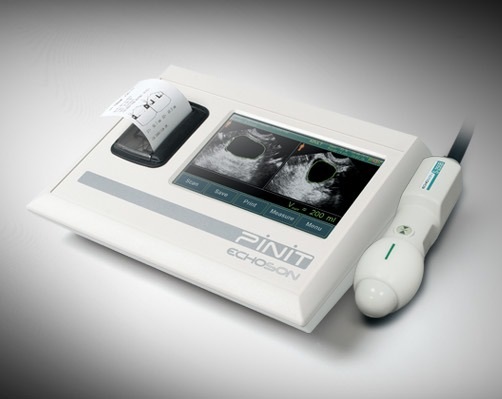
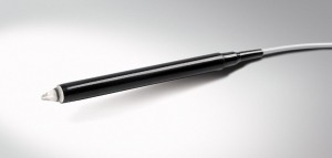
CCT- PACHYMETER (P-SCAN) –  Ocular cornea biometrics 
Up-to-date ultrasonic pachymeter  used to measure the thickness of the eye’s cornea (glaucoma screening and refractive surgery),  with a very high sampling frequency –400MHz  (a significant increase in measurement accuracy – ≤ 2 μm). Corneal pachymetry is an important test in the early detection of glaucoma (central corneal thickness CCT), and it help also surgeons by providing graphical surgical plans to eliminate corneal astigmatism.

PIROP user’s interface based on touch screen technology makes operation easy and user-friendly. The touch screen can be also operated not only with fingers, but also using the enclosed stylus to keep it clean.


Specifications
A-SCAN
Complete and quick biometry :
– eye axial length (AXL)
– anterior chamber depth (AC)
– lens thickness (LENS)
– vitreous length (VITR)
Measurement of all eye types: normal, cataract, dense cataract, aphakic,silicone oil vitreous, pseudoaphakic (PMMA, ACRYLATE, SILICONE)
OL six formulas: SRK II, SRK T, Holladay, Hoffer-Q, Binkhorst II, Haggis
Post refractive formulas: Double-K SRKT, Latkany / Flat-K SRKT/, Latkany /avg-K SRKT/, Masket
IOL power calculation comparison (for different lenses and calculation methods)
Contact and immersion methods (the immersion tube attached)
Probe with central light beam for correct eye positioning
Automatic TGC adjustment;GAIN adjustment up to 110dB
Memory of 20 scans with measurements for further analysis
Calculations of means and standard deviations
Automatic control of measurement scatters (with manual correction)
Customizable for 10 users, 10 profiles for each.
Adjusting and testing via attached calibrator
Probe frequency – 12 MHz
Range of scan 45 mm / 2500 points per line
Clinical resolution 0.1 mm
Electronic resolution –/+ 0.01 mm
POWER SUPPLY
Power supply from external AC/DC power adapter:
– input: 100-230V AC 50/60Hz / max 0.7A
– output: +12V DC / 2 A
Power consumption (12 DC supply) – ca 12 W
Electrical safety standards
– Medical device Class IIa complies with MDD 93/42 EEC
– Scanner complies with requirements for Class II devices of EN/IEC 60601-1
B-SCAN
Probe frequency: 12 .. 15 MHz
Scanning angle : 55ᵒͦ
Range of scan: 20 .. 60 mm
Gain/dymamics up to 110dB / 90dB
TGC – zone gain adjustment in selected eye
256 Levels Gray Scale
Axial clinical resolution: 0.12 mm
Lateral clinical resolution : 0.3 mm
Display modes : B, B+A, B+B
A-mode for selected line CV (in Run and Freeze modes)
Dynamic Range Correction
Image cache memory M1..M4 independent for right and left eyes
Zoom function (x2) in Run and Freeze modes
Digital Gain Correction in Freeze mode
Cineloop function (about 6 second) independent for left/right eyes
Distances measurements with ultrasound velocity correction
Area and angle measurements
Pointers, comments entered by the user
Composite video output for videoprinter
CCT – PACHYMETER
Quick thickness measurements for all corneal types
– the measurement of central corneal thickness (CCT)
– thickness measurement at arbitrarily selected points
– automatic and manual measurements
– calculation of mean from measurements
– standard deviation of the measured thickness
– ability for rejecting inaccurate measurements
Very high level of accuracy and repeatability of all measurements:
– sophisticated algorithms for the accuracy improvement
– very high frequency for measuring purpose- 400 MHz
– high averaging ratio
– 512 automatic measurements cycles
Intra-ocular pressure (IOP) calculations with measuring and/or manual correction
Ten IOP correction formulas : Kohlhas/Shah, Argus et al. Whitarce et al, Doughty, Ehlers, Dresden, Stodmeister, custom-own
BIAS percentage deviation of the measured thickness from 60% to 130%
TEST function for automatic checking of operation correctness (without using external phantoms or patterns)
Nine defined maps of corneal thickness with the number of points: 1, 5, 9, 13, 21, 25, 1 MULTI, 5 MULTI, 9 MULTI (central, paracentral, peripheral, parietal, mixed)
20 MHz probe operating frequency
Default sound velocity – 1640 m/s ; range from 1400 to 2000 m/s
Measurement range from 220 μm to 1100 μm
Measuring accuracy ≤ 2 μm
Resolution 1 μm
GENERAL PROPERTIES
Touch Screen
– easy, ergonomic and user-friendly operation via displayed menu
– virtual keyboard for entering patient data, etc.
Functional knob (gain control, review results, cine etc.)
Color LCD 7” display
Internal patient’s databas
Ten (10) user profiles
Support for barcode reader
Possibility of connecting an external alphanumeric keyboard
Recording of images in internal and USB-Pendrive memory.
Internal thermal small printer ( for A-scan and P-scan reports )
Composite-video output PAL (for videoprinter or video monitor )
Support of multiple languages
Easy firmware upgrade via USB port
PIROP-PC software (optional)
a special software that enables management of the patient database and measurements (images) obtained from PIROP scanner.
it can be installed in any external PC computer, tablet etc. operated by Windows.
exchange of images and patient data between a scanner and the PIROP-PC application is done using an USB memory or via a direct USB connection.
possibility to print on any printer the special report of exams
additional option – possibility to use the standard DICOM 3.0 to communicate with PACS/RIS systems (Store, Print, Worklist).
WEIGHT AND DIMENSIONS
Dimensions (with built-in printer): 290 x 205 x 85 mm
Weight (with built-in printer): 1.5 kg
Probes
OA12 A-scan
Frequency: 20 MHz
Application: biometry and lens power calculation for intraocular implants.
OB12 B-Scan
Frequency: 12/15 MHz
Application: imaging of interior of an eyeball, retina, optical nerve ..
OP20 Pachymetr
Frequency: 20 MHz
Application: measurements of thickness for all corneal types
E - brochures
Scanner images
Images gallery